BeambePrep / MDCAT & NUMS Syllabus Biology • Chapter 1: Homeostasis
Excretory System & Renal Function

Homeostasis

Chapter Contents & Quick Jump 6 Sections Click to expand

Core Principles of Homeostasis

Homeostasis is the dynamic maintenance of a relatively stable internal environment despite continuous external disturbances. It shields cellular biochemistry from catastrophic environmental fluctuations.

The internal physical state is anchored around a precise set point. Human homeostasis prioritizes three physiological variables:

  • Water Volume and osmotic equilibrium.
  • Dissolved Solute Concentrations (electrolytes like sodium, potassium, and chloride).
  • Core Body Temperature anchored around 37 C.

Regulation operates via feedback control loops comprising three biological components:

  1. Receptors (Sensors): Detect internal or external environmental deviations.
  2. Control Center (Integrator): Processes sensory input, compares it against the set point, and signals the response pathway.
  3. Effectors: Muscles or glands that execute corrective actions to restore equilibrium.

Three Pillars of Excretory Homeostasis

  • Osmoregulation: Active control of internal solute concentrations and water balance across biological membranes.
  • Excretion: Removal of toxic nitrogenous metabolic byproducts, specifically ammonia, urea, and uric acid.
  • Thermoregulation: Maintenance of internal core temperature within functional physiological limits.
Crucial Physiological Boundary
Do kidneys directly coordinate human thermoregulatory sweating and shivering?
No! The hypothalamic thermostat regulates body temperature. The kidneys govern blood volume, electrolyte balance, and nitrogenous urea elimination.
Provincial Board Variance
BTB: BTB defines the normal baseline value of any homeostatic parameter as the set point.
STB: STB specifies that excretion simultaneously maintains osmotic balance, ionic balance, and acid-base equilibrium.
Gross Kidney Anatomy & Urinary Pathways

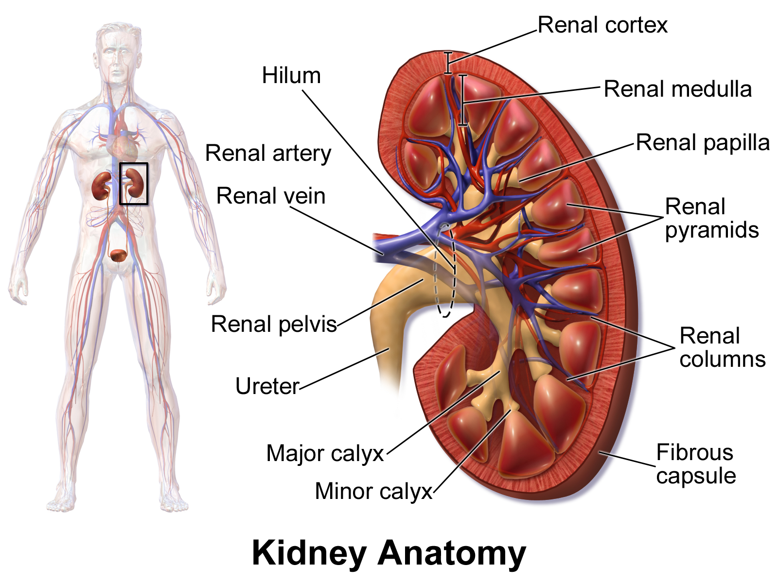
Human kidneys are bilateral, reddish-brown, bean-shaped organs positioned retroperitoneally against the posterior abdominal wall, situated between the T12 and L3 vertebrae.

The right kidney sits slightly lower than the left kidney to accommodate the large right lobe of the liver. Each kidney displays a medial concave indentation termed the hilus, through which the renal artery, renal vein, lymphatic vessels, nerves, and ureter pass.

Gross renal parenchyma divides into three concentric structural regions:

  • Renal Cortex: The outer reddish granular zone containing all renal corpuscles (glomeruli and Bowman capsules) as well as cortical convoluted tubules.
  • Renal Medulla: The inner striated zone organized into 8 to 18 conical renal pyramids whose apical papillae project toward the calyces.
  • Renal Pelvis: The central funnel-shaped chamber that pools urine from major calyces and conveys it into the ureter.
Provincial Board Variance
KTB: KTB notes human kidneys measure about 12 cm long, 6 cm wide, 4 cm thick, and weigh approximately 150 g.
BTB: BTB notes kidneys are each approximately 10 cm in length.
STB: STB notes kidneys are 4 to 5 inches long.
BLAUSEN MEDICAL CC-BY
Plate 1.1 · Gross Kidney Anatomy, Zonal Division, and Vascular Entry

Nephron Structure & Microvascular Organization

The nephron is the structural and functional filtration unit of the human kidney. Each adult kidney contains approximately 1.0 to 1.2 million nephrons.

A nephron consists of two major functional divisions:

  1. Renal Corpuscle: Comprising the glomerular capillary tuft encased within the double-walled Bowman capsule.
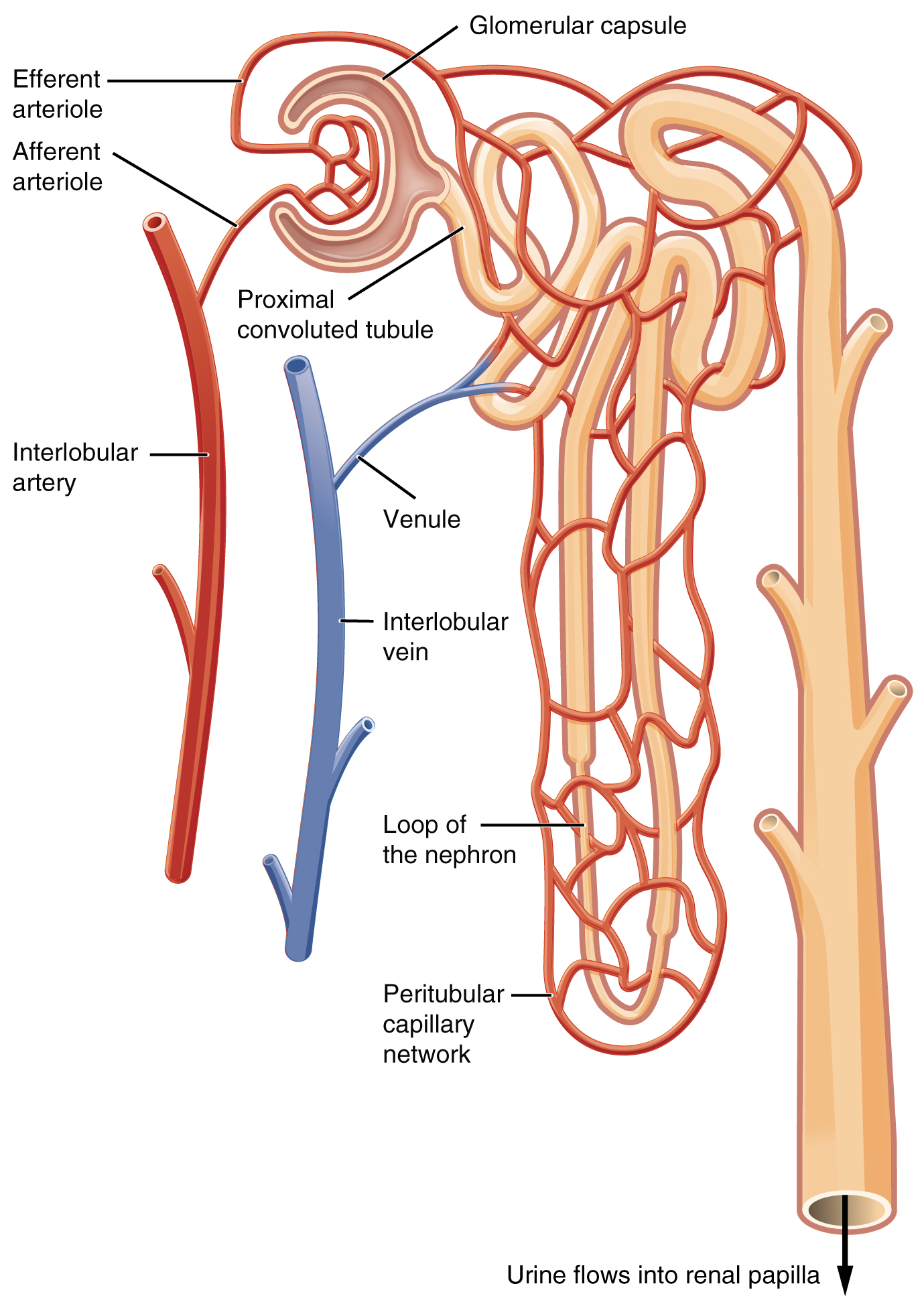
  2. Renal Tubule: Consisting of the Proximal Convoluted Tubule (PCT), the hairpin Loop of Henle, the Distal Convoluted Tubule (DCT), and terminal collecting ducts.

Nephrons fall into two distinct anatomical classes:

  • Cortical Nephrons (~70 to 80%): Renal corpuscles lie in the outer cortex. Loops of Henle are short and penetrate only into the outer medulla. Their primary role is bulk filtration and standard reabsorption under normal hydration.
  • Juxtamedullary Nephrons (~20 to 30%): Renal corpuscles lie near the corticomedullary junction. Loops of Henle plunge deep into the hypertonic inner medulla, accompanied by specialized hairpin capillary loops called vasa recta. They generate the osmotic gradient essential for concentrating urine during water conservation.

The renal circulation features a unique portal-like arrangement:

The glomerulus is positioned between two resistance arterioles rather than an arteriole and a venule. The wider diameter of the afferent arteriole paired with the narrower efferent arteriole creates elevated hydrostatic filtration pressure within the capillary bed.

Provincial Board Variance
KTB: Cortical nephrons comprise 70 to 80 percent, juxtamedullary nephrons comprise 20 to 30 percent.
BTB: Cortical nephrons account for 80 to 85 percent, juxtamedullary nephrons account for 15 to 20 percent.
OPENSTAX CC-BY 4.0
Plate 1.2 · Nephron Microvasculature and Arteriolar Resistance Bed

Urine Formation: Filtration, Reabsorption, and Secretion

Urine production involves three sequential, highly coordinated physiological processes:

1. Glomerular Ultrafiltration

Non-selective bulk filtration powered by capillary hydrostatic pressure (~55 to 60 mmHg). Fluid and small solutes cross the three-layered filtration barrier (fenestrated capillary endothelium with pores of 70 nm, basement membrane, and podocyte filtration slits).

Blood cells and large plasma proteins like albumin are excluded. Normal Glomerular Filtration Rate (GFR) is 125 ml/min, yielding 180 Liters of filtrate every 24 hours.

2. Selective Tubular Reabsorption

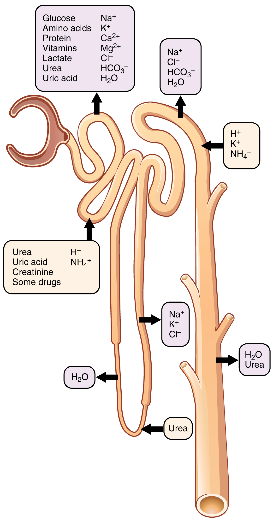
Over 99.5% of filtered water, ions, and nutrients are returned to the peritubular capillary blood.

The Proximal Convoluted Tubule (PCT) reabsorbs:

  • 100% of filtered glucose and amino acids via sodium-dependent secondary active transport.
  • Approximately 65 to 80% of filtered sodium chloride and water.

3. Tubular Secretion

The selective transfer of substances from peritubular capillaries across tubular epithelium directly into the lumen:

  • Secretion of hydrogen ions ($H^+$) and ammonium ($NH_4^+$) directly controls systemic blood pH.
  • Secretion of potassium ($K^+$), creatinine, and medicinal metabolites like penicillin.
Provincial Board Variance
STB: STB notes the kidneys filter 180 liters of blood in 24 hours to generate 2.5 liters of urine.
PTB: PTB and BTB note kidneys filter 180 liters of blood to generate 1.0 to 1.5 liters of normal urine.
OPENSTAX CC-BY 4.0
Plate 1.3 · Selective Tubular Reabsorption and Secretion Sites along the Nephron

Osmoregulation & The Counter-Current Multiplier

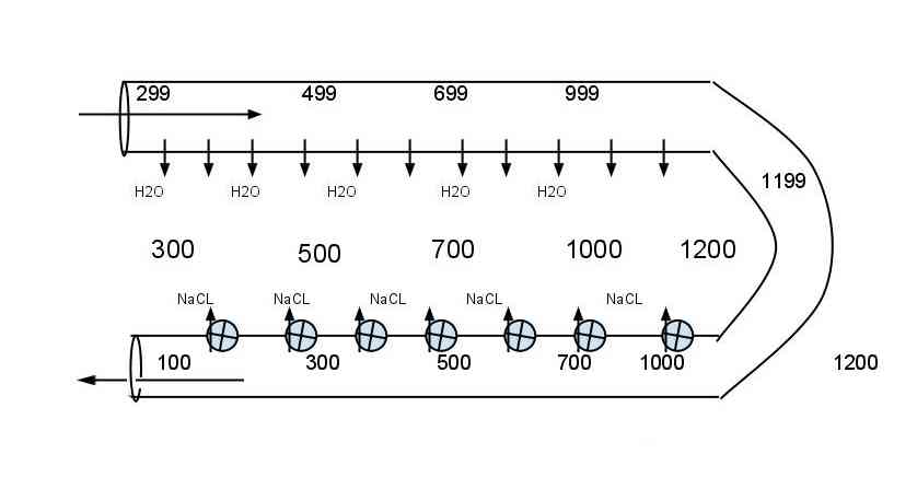
The kidney regulates blood osmolarity by creating a steep osmotic gradient in the medullary interstitium, rising from 300 mOsm/L in the cortex to 1200 mOsm/L in the deep renal papillae.

This osmotic gradient is built by the Counter-Current Multiplier within the Loop of Henle:

  • Descending Limb: Highly permeable to water via aquaporin channels, but completely impermeable to solutes ($NaCl$). As tubular fluid descends, water exits into the hypertonic medulla via osmosis, concentrating tubular fluid up to 1200 mOsm/L at the hairpin turn.
  • Ascending Limb: Completely impermeable to water. The thin segment permits passive $NaCl$ diffusion, while the thick ascending limb actively pumps $Na^+$ and $Cl^-$ out into the medullary interstitium via $Na^+/K^+/2Cl^-$ cotransporters. Fluid leaving the loop into the DCT becomes hypotonic (~100 to 200 mOsm/L).

The Vasa Recta act as Counter-Current Exchangers. Their hairpin design allows blood to flow passively through the medulla, taking up reabsorbed water while leaving the interstitial salt gradient undisturbed.

Hormonal regulation completes the osmoregulatory circuit:

  • Antidiuretic Hormone (ADH / Vasopressin): Released by posterior pituitary during dehydration. Stimulates aquaporin-2 insertion into collecting duct luminal membranes, yielding concentrated, low-volume urine.
  • Aldosterone: Secreted by adrenal cortex in response to renin-angiotensin activation. Promotes active $Na^+$ reabsorption and $K^+$ excretion in the distal tubule, defending extracellular fluid volume.
WIKIMEDIA CC-BY-SA
Plate 1.4 · The Counter-Current Multiplier and Interstitial Osmotic Gradient

Renal Pathologies & Clinical Interventions

Kidney Stones (Renal Calculi)

Kidney stones precipitate when mineral salts supersaturate in urine:

  • Calcium Oxalate & Phosphate: Account for 70% of all renal calculi. Hypercalcemia and diets rich in oxalates (green leafy vegetables, tomatoes) accelerate crystallization.
  • Struvite (Infection Stones): Account for 15 to 20%. Caused by urea-splitting bacterial urinary tract infections.
  • Uric Acid Stones: Account for 5 to 10%. Linked to hyperuricemia and purine metabolism disorders.
  • Cystine Stones: Rare 1 to 3%, caused by inherited amino acid transporter defects.

Non-invasive stone removal uses Extracorporeal Shock Wave Lithotripsy (ESWL), which pulverizes calculi into sandy fragments using high-energy acoustic shock waves.

Renal Failure & Replacement Therapies

Renal failure represents progressive loss of filtration capacity, accumulating urea and metabolic toxins (a condition termed uremia).

  • Hemodialysis: Blood is routed through an artificial dialyzer cartridge where dialysate fluid flows counter-current to blood across cellophane membranes, clearing urea by diffusion.
  • Peritoneal Dialysis: Dialysis fluid is introduced into the patient's abdominal cavity using the vascularized peritoneum as the natural semi-permeable filtration membrane.
  • Kidney Transplantation: The definitive clinical replacement for End-Stage Renal Disease (ESRD).
Provincial Board Variance
KTB: When kidney function falls below 10% of normal, dialysis or kidney transplant is required.
STB: STB notes kidney transplantation is indicated when kidneys have lost about 90% of their functional ability.
High-Yield Past Paper Hits
The definitive long-term clinical therapy for end-stage renal failure is donor kidney transplantation. NUMS 2024
In peritoneal dialysis, dialysate fluid is introduced directly into the peritoneal abdominal cavity. UHS 2023
Uric acid stones account for approximately 5 to 10 percent of total renal stones. KMU 2022

Ready to test your active recall on Homeostasis?

Master this chapter with 185 FSRS flashcards in the Pulse Deck, and practice 112 verified past paper questions and topical MCQs in the BeambePrep Swarm Engine.

MDCAT & NUMS Syllabus Tags
#Homeostasis #Nephron #UrineFormation #CounterCurrentMultiplier #MDCATBiology #PMDCSyllabus #NUMS2026 #PTB #STB #KTB #BTB #FederalBoard #ActiveRecall